File:Flagellata 1.png

From Wikimedia Incubator

Original file(990 × 2,136 pixels, file size: 147 KB, MIME type: image/png)

This file is from Wikimedia Commons and may be used by other projects. The description on its file description page there is shown below.

Summary

Description
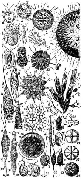
English: * 1. — Chlamydomonas pulvisculus, Ehr. (Chlamydomonadidae) free-swimming individual.
    • a = nucleus.
    • b = contractile vacuole.
    • c = starch corpuscle.
    • d = cellulose investment.
    • e = stigma (eye-spot).
  • 2. Resting stage of the same, with fourfold division of the cell-contents. Letters as before.
  • 3. Breaking up of the cell-contents into minute biflagellate swarm-spores, which escape, and whose history is not further known.
  • 4. Syncrypta volvox, Ehr. (Chrysomonadidae). A colony enclosed by a common gelatinous test c.
    • a = stigma.
    • b = vacuole (non-contractile).
  • 5. Uroglena volvox, Ehr. (Chrysomonadidae). Half of a large colony, the flagellates embedded in a common jelly.
  • 6. Chlorogonium euchlorum, Ehr. (Chlamydomonadidae).
    • a = nucleus.
    • b = contractile vacuole.
    • c = starch grain.
    • d = eye-spot.
  • 7. Chlorogonium euchlorum, Ehr. (Chlamydomonadidae). Copulation of two liberated microgonidia.
    • a = nucleus.
    • b = contractile vacuole.
    • d = eye-spot (so-called).
  • 8. Colony of Dinobryon sertularia, Ehr. (Chrysomonadidae).
  • 9. Haematococcus palustris, Girod (= Chlamydococcus, Braun, Protococcus, Cohn), one of the Chrysomonadidae; ordinary individual with widely separated test.
    • a = nucleus.
    • b = contractile vacuole.
    • c = amylon nucleus (pyrenoid).
  • 10. Dividing resting stage of the same, with eight fission products in the common test e.
  • 11. A microgonidium of the same.
  • 12. Phalansterium consociatum, Cienk. (Choanoflagellata); × 325. Disk-like colony.
  • 13. Euglena viridis, Ehr.; × 300 (Euglenidae).
    • a = pigment spot (stigma).
    • b = clear space.
    • c = paramylum granules.
    • d = chromatophor (endochrome plate).
  • 14. Gonium pectorale, O. F. Müller (Volvocineae). Colony seen from the flat side; × 300.
    • a = nucleus.
    • b = contractile vacuole.
    • c = amylon nucleus.
  • 15. Dinobryon sertularia, Ehr. (Chrysomonadidae).
    • a = nucleus.
    • b = contractile vacuole.
    • c = amylon nucleus.
    • d = free colourless flagellates, probably not belonging to Dinobryon.
    • e = stigma (eye-spot).
    • f = chromatophors.
  • 16. Peranema trichophorum, Ehr. (Peranemidae), creeping individual seen from the back; × 140.
    • c = pharynx.
    • d = mouth.
  • 17. Anterior end of Euglena acus, Ehr., in profile.
    • a = mouth.
    • b = vacuoles.
    • c = pharynx.
    • d = stigma (eye-spot).
    • e = paramylum-body.
    • f = chlorophyll corpuscles.
  • 18. Part of the surface of a colony of Volvox globator, L. (Volvocidae), showing the intercellular connective fibrils.
    • a = nucleus.
    • b = contractile vacuole.
    • c = starch granule.
  • 19. Two microgametes (spermatozoa) of Volvox globator, L.
    • a = nucleus.
    • b = contractile vacuole.
  • 20. Ripe asexually produced daughter-individual of Volvox minor, Stein, still enclosed in the cyst of the partheno-gonidium.
    • a = young, partheno-gonidia.
  • 21, 22. Trypanosoma sanguinis, Gruby (Haematoflagellates), from the blood of Rana esculenta.
    • a = nucleus; × 500.
  • 23—26. Reproduction of Bodo caudatus, Duj. (Bodonidae), after Dallinger and Drysdale:—23, fusion of several individuals (plasmodium); 24, encysted fusion-product dividing into four; 25, later into eight; 26, cyst filled with swarm-spores.
  • 27. Distigma proteus, Ehbg., O.F. Müller (Euglenidae); × 440. Individual with the two flagella, and strongly contracting hinder region of the body.
  • 28. The same devoid of flagella.
    • c, c = the two dark pigment spots (so-called eyes) near the mouth.
  • 29. Oicomonas termo (Monas termo) Ehr. (one of the Oicomonadidae).
    • c = food-ingesting vacuole.
    • d = food-particle; × 440.
  • 30. The food-particle d has now been ingested by the vacuole.
  • 31. Oicomonas mutabilis, Kent (Oicomonadidae), with adherent stalk.
    • a = nucleus.
    • b = contractile vacuole.
    • c = food-particle in food vacuole.
  • 32, 33. Cercomonas crassicauda, Duj. (Oicomonadidae), showing two conditions of the pseudo-podium-protruding tail.
    • a = nucleus.
    • b = contractile vacuoles.
    • c = mouth.
Source Encyclopædia Britannica
Author Encyclopædia Britannica

Licensing

Public domain This image comes from the 13th edition of the Encyclopædia Britannica or earlier. The copyrights for that book have expired in the United States because the book was first published in the US with the publication occurring before January 1, 1929. As such, this image is in the public domain in the United States.

Captions

Add a one-line explanation of what this file represents

In dieser Datei abgebildete Objekte

depicts

image/png

File history

Click on a date/time to view the file as it appeared at that time.

Date/TimeThumbnailDimensionsUserComment
current13:59, 12 April 2006Thumbnail for version as of 13:59, 12 April 2006990 × 2,136 (147 KB)Jjbeard{{PD-Britannica}} Category:Images from Encyclopædia Britannica This image has an extensive caption. Please refer to the EB1911 article for the text of this caption.

There are no pages that use this file.

Global file usage

The following other wikis use this file: